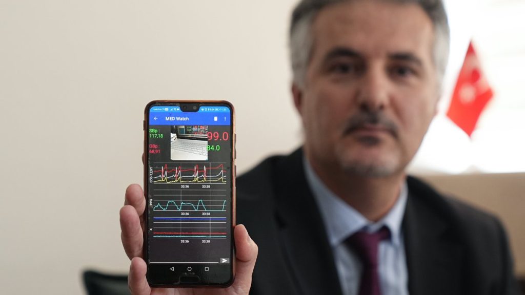
Scientists at Ankara University (AU) have developed an artificial intelligence-supported application that could enable the early detection of lung cancer by analyzing people’s speech patterns.
The research team, led by Assoc. Prof. Dr. Haydar Ankışhan from AU’s Stem Cell Institute, collaborated across disciplines to create the application, aiming to diagnose lung cancer, a disease typically detected at later stages, much earlier.
Speaking at a press conference held at AU’s Ibn-i Sina Hospital, Ankışhan explained that the project targets the structural changes in the voice that may reflect underlying alterations in lung anatomy and function.
“In our study, we considered the structure of the voice, the anatomical structure of the lungs, and the circulatory system. We proposed that the voice could provide information about lung cancer,” Ankışhan said.
“After approximately a year and a half of research, we achieved promising results, finding that lung cancer could be detected at the first stage with over 90% accuracy,” he added.
The application works by recording voices in a natural environment and analyzing them using advanced signal processing and AI techniques developed by the research team. By training the AI with these data, researchers can distinguish between healthy individuals and those with early-stage lung cancer.
Assoc. Prof. Dr. Bülent Mustafa Yenigün, a faculty member at AU’s Faculty of Medicine and another key contributor to the project, stressed the importance of early detection in the successful treatment of lung cancer.
He highlighted that the team sought a low-cost, non-invasive method that could reduce patients’ exposure to harmful imaging procedures like x-rays.
The initial study was conducted with a group of 50 lung cancer patients and 50 healthy individuals. Each participant read a prepared two-minute speech, and all recordings were made under identical conditions. Comparisons between the groups yielded a diagnostic accuracy rate of up to 92%.
However, Yenigün pointed out that the sample size remains small. “In the future, when thousands of patient recordings are gathered, we believe the results will be even better, with a much lower margin of error,” he said.
Explaining the science behind the method, Yenigün noted that masses in the lungs alter airflow and voice resonance. “Our application detects these changes in sound frequency and resonance, signaling that a pathological mass could be present,” he explained.
Regarding the timeline for broader use, the researchers estimate that the application could be integrated into screening programs within two to three years, provided enough data is collected and legal approvals are secured. If these steps are completed quickly, the application could become available in as little as one to two years.


